Bap1 Mtap Mesothelioma - Jcm Free Full Text When The Diagnosis Of Mesothelioma Challenges Textbooks And Guidelines Html - bap1 immunohistochemistry and p16 fluorescence in situ hybridization (fish) have recently been reported as reliable markers of malignancy in biopsies of mesothelioma.
Bap1 Mtap Mesothelioma - Jcm Free Full Text When The Diagnosis Of Mesothelioma Challenges Textbooks And Guidelines Html - bap1 immunohistochemistry and p16 fluorescence in situ hybridization (fish) have recently been reported as reliable markers of malignancy in biopsies of mesothelioma.. Huang jt, liu yj, wang j, xu zg, yang y, shen f, et al. Homozygous deletion of cdkn2a detected by fluorescence in situ hybridization is a diagnostic marker for malignancy in mesothelioma. Cytologically, the tubules are lined by bland cuboidal epithelium. mtap is located adjacent to cdkn2a and is codeleted in 90% of mesothelioma tumors with a cdkn2a homozygous deletion. mtap a new added marker for diagnosis of mesothelioma in cytology #tis @fcschmitt #molcytopath20 @ashishc97225686 @sinchita_roy (@swikrityumd) 9 months ago.
These are calcifications with an unusual and pretty lamellar pattern. A combination of ezh2, bap1 ihc, and p16 fish showed a sensitivity of 89.5%; Comparison with 9p21 fish and bap1 immunohistochemistry. Comparison with 9p21 fish and bap1 immunohistochemistry. The body's mtap enzyme was lost to only 23 percent of epithelioid tests and 24 percent of sarcomatoid mesothelioma tests.
Cdkn2a Copy Number And P16 Expression In Malignant Pleural Mesothelioma In Relation To Asbestos Exposure Bmc Cancer Full Text from media.springernature.com Comparison with 9p21 fish and bap1 immunohistochemistry. Figure 2 bap1 and mtap immunohistochemistry in mesothelioma pleural effusion cytology. Malignant pleural mesothelioma (mpm) is a highly lethal cancer of the lining of the chest cavity. Loss of nuclear bap1 staining in a population of tumor cells defined by ihc as mesothelial in origin is diagnostic of malignant mesothelioma. Homozygous deletion of cdkn2a detected by fluorescence in situ hybridization is a diagnostic marker for malignancy in mesothelioma. Both of these proteins are normally found in tissues throughout the body. Immunohistochemical detection of mtap and bap1 protein loss for mesothelioma diagnosis: Mechanisms through which bap1 inactivation occurs include mutation, copy number loss, or translocations 4.
A firm diagnosis of malignant mesothelioma needs to be made before an effective treatment plan can be put into place, and after that initial diagnosis is made surgeons need to be able to distinguish between benign and malignant tissue.
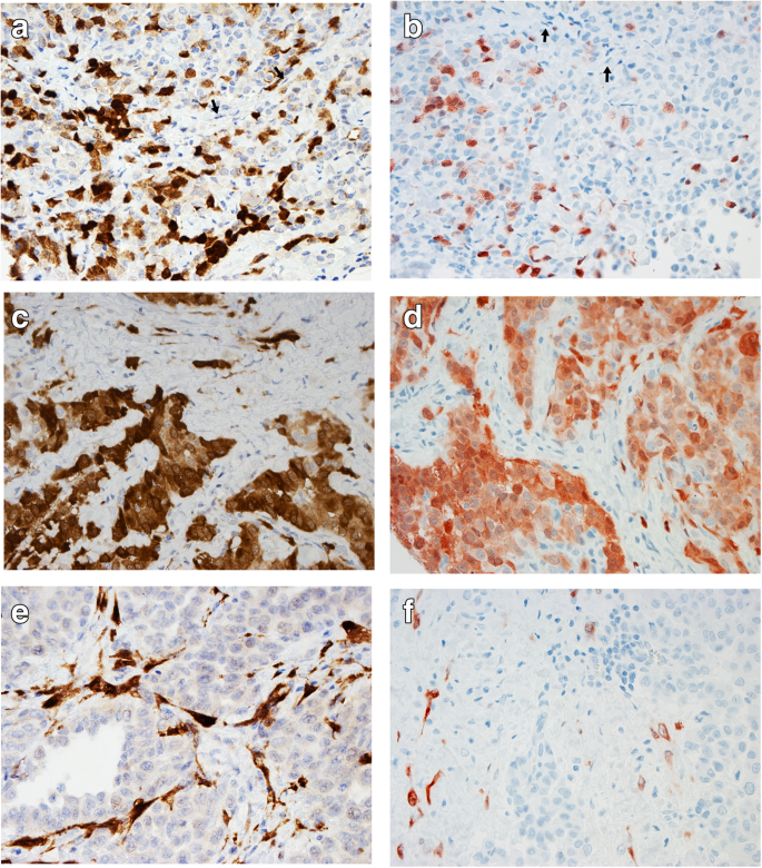
To determine whether these markers, singly or in combination, might also be useful in effusion cytology specimens, we examined 15 biopsies. Three cases were not further investigated based on patient and clinician decision. Suppression of ezh2 using rna interference was found to decrease the cancerogeneity of malignant mesothelioma cells. However, they are not typically seen in mesothelioma. Su c, chang y, chan y, et al: Immunohistochemical detection of mtap and bap1 protein loss for mesothelioma analysis. Methylthioadenosine phosphorylase (mtap) is a 9p21.3 related protein involved in purine metabolism that plays a role in salvage of adenosine and methionine and is expressed in mesothelial cells. Immunohistochemical detection of mtap and bap1 protein loss for mesothelioma diagnosis: Loss of nuclear bap1 staining in a population of tumor cells defined by ihc as mesothelial in origin is diagnostic of malignant mesothelioma. They found that all markers had a speci ficity equal to 100%, whereas sensitivities of ezh2, bap1, mtap ihc, and p16 fish were equal to 44.7%, 52.6%, 47.4%, and 65.8%, respectively. Immunohistochemical detection of mtap and bap1 protein loss for mesothelioma diagnosis: At the interface of more dense fibrous and more reactive inflammatory fibrous tissue there is a linear proliferation of tubules with focal papillary formation. (a) h&e stained section prepared from cell block shows moderate to high cellularity consists of predominantly mesothelial cells, arranged in simple clusters and papillaroid groups.
Pleural effusions are among the first clinical manifestations of malignant pleural mesothelioma (mpm) and often constitute the only available material for diagnosis. Mechanisms through which bap1 inactivation occurs include mutation, copy number loss, or translocations 4. Sarcomatoid mesothelioma presents additional difficulties, because the tumors can resemble benign (noncancerous) conditions or other types of cancer. Malignant mesothelioma, reactive mesothelial proliferation, bap1, mtap, cdkn2a, malignant mesothelioma in situ search for similar articles you may search for similar articles that contain these same keywords or you may modify the keyword list to augment your search. Methylthioadenosine phosphorylase (mtap) is a 9p21.3 related protein involved in purine metabolism that plays a role in salvage of adenosine and methionine and is expressed in mesothelial cells.
Heg1 Bap1 And Mtap Are Useful In Cytologic Diagnosis Of Malignant Mesothelioma With Effusion Hiroshima 2021 Diagnostic Cytopathology Wiley Online Library from onlinelibrary.wiley.com None of the mesothelioma cases showed homozygous nf2 deletion. mtap is an independent prognosis marker and the concordant loss of mtap and p16 expression. Sarcomatoid mesothelioma presents additional difficulties, because the tumors can resemble benign (noncancerous) conditions or other types of cancer. These are calcifications with an unusual and pretty lamellar pattern. A firm diagnosis of malignant mesothelioma needs to be made before an effective treatment plan can be put into place, and after that initial diagnosis is made surgeons need to be able to distinguish between benign and malignant tissue. To expand our understanding of mpm, we conducted a comprehensive integrated genomic study, including the most detailed analysis of bap1 alterations to date. Malignant pleural mesothelioma diagnostics immunohistochemistry fluorescence in situ hybridization. At the interface of more dense fibrous and more reactive inflammatory fibrous tissue there is a linear proliferation of tubules with focal papillary formation.
Loss of nuclear bap1 staining in a population of tumor cells defined by ihc as mesothelial in origin is diagnostic of malignant mesothelioma.
Huang jt, liu yj, wang j, xu zg, yang y, shen f, et al. A combination of mtap and bap1 immunohistochemistry in pleural effusion cytology for the diagnosis of mesothelioma. Hemizygous nf2 loss (chromosome 22 monosomy or hemizygous deletion) was detected in 25 of 47 (53.2%) mesothelioma cases. Immunohistochemical detection of mtap and bap1 protein loss for mesothelioma diagnosis: Deletion of the 9p21.3 chromosome region and loss of mtap (or bcra1 associated protein 1: Immunohistochemical detection of mtap and bap1 protein loss for mesothelioma analysis. Histologic diagnosis of malignant pleural mesothelioma (mpm) is not always straightforward. Although an mpm diagnosis can be reliable on cytology, the reported sensitivity is low (30% to 75%). The former is used for amp and the latter for methionine synthesis. In clinic practice, highly expressed ezh2 and deletion of bap1 and mtap can be detected by immunohistochemistry and may be used to distingush mpm from mesothelial hyperplasia. Highly expressed ezh2 in combination with bap1 and mtap loss, as detected by immunohistochemistry, is useful for differentiating malignant pleural mesothelioma from reactive mesothelial hyperplasia. Malignant pleural mesothelioma diagnostics immunohistochemistry fluorescence in situ hybridization. Loss of nuclear bap1 staining in malignant mesothelioma.
Loss of nuclear bap1 staining in a population of tumor cells defined by ihc as mesothelial in origin is diagnostic of malignant mesothelioma. Pleural effusions are among the first clinical manifestations of malignant pleural mesothelioma (mpm) and often constitute the only available material for diagnosis. Significant progress in the diagnosis and classification of pleural mesothelioma has been made in the past 5 years. Although an mpm diagnosis can be reliable on cytology, the reported sensitivity is low (30% to 75%). mtap is an independent prognosis marker and the concordant loss of mtap and p16 expression.
Highly Expressed Imp3 And Glut 1 In Combination With The Loss Of Bap1 Expression Is Useful For Differentiating Malignant Mesothelioma From Reactive Mesothelial Hyperplasia Research Square from assets.researchsquare.com Immunohistochemical detection of mtap and bap1 protein loss for mesothelioma diagnosis: mtap deficiency leads to a dependency on de novo purine synthesis. bap1 immunohistochemistry and p16 fluorescence in situ hybridization (fish) have recently been reported as reliable markers of malignancy in biopsies of mesothelioma. Hida t, hamasaki m, matsumoto s, et al: (13.) bianchi a, mitsunaga s, cheng j, et al. Lung most cancers is most frequently attributable to smoking whereas mesothelioma is most frequently attributable to. Therefore, this stain is not useful to distinguish between these two malignancies. At the interface of more dense fibrous and more reactive inflammatory fibrous tissue there is a linear proliferation of tubules with focal papillary formation.
Loss of nuclear bap1 staining in a population of tumor cells defined by ihc as mesothelial in origin is diagnostic of malignant mesothelioma.
Immunohistochemical detection of mtap and bap1 protein loss for mesothelioma diagnosis: mtap a new added marker for diagnosis of mesothelioma in cytology #tis @fcschmitt #molcytopath20 @ashishc97225686 @sinchita_roy (@swikrityumd) 9 months ago. bap1) protein expression is a reliable marker for malignant mesothelioma diagnosis. Deletion of the 9p21.3 chromosome region and loss of mtap (or bcra1 associated protein 1: Immunohistochemical detection of mtap and bap1 protein loss for mesothelioma analysis. Although an mpm diagnosis can be reliable on cytology, the reported sensitivity is low (30% to 75%). Lung most cancers is most frequently attributable to smoking whereas mesothelioma is most frequently attributable to. bap1 ihc, mtap ihc, and cdkn2a fish, and we suggest that these should be the first approach to the problem. Hida t, hamasaki m, matsumoto s, et al: mtap is an independent prognosis marker and the concordant loss of mtap and p16 expression. This rate is similar to what is observed in sarcomatoid mesothelioma (61%). High frequency of inactivating mutations in the neurofibromatosis type 2 gene (nf2. bap1 (brca1 associated protein 1) 14d.


0 Comments